TorchXRayVision is an open source software library for working with chest X-ray datasets and deep learning models. It provides a common interface and common pre-processing chain for a wide set of publicly available chest X-ray datasets. In addition, a number of classification and representation learning models with different architectures, trained on different data combinations, are available through the library to serve as baselines or feature extractors.
Gifsplanation via Latent Shift: A Simple Autoencoder Approach to Counterfactual Generation for Chest X-rays
Motivation: Traditional image attribution methods struggle to satisfactorily explain predictions of neural networks. Prediction explanation is important, especially in medical imaging, for avoiding the unintended consequences of deploying AI systems when false positive predictions can impact patient care. Thus, there is a pressing need to develop improved models for model explainability and introspection.
Specific problem: A new approach is to transform input images to increase or decrease features which cause the prediction. However, current approaches are difficult to implement as they are monolithic or rely on GANs. These hurdles prevent wide adoption.
Our approach: Given an arbitrary classifier, we propose a simple autoencoder and gradient update (Latent Shift) that can transform the latent representation of a specific input image to exaggerate or curtail the features used for prediction. We use this method to study chest X-ray classifiers and evaluate their performance. We conduct a reader study with two radiologists assessing 240 chest X-ray predictions to identify which ones are false positives (half are) using traditional attribution maps or our proposed method.
Results: We found low overlap with ground truth pathology masks for models with reasonably high accuracy. However, the results from our reader study indicate that these models are generally looking at the correct features.
We also found that the Latent Shift explanation allows a user to have more confidence in true positive predictions compared to traditional approaches (0.15±0.95 in a 5 point scale with p=0.01) with only a small increase in false positive predictions (0.04±1.06 with p=0.57).
Saliency is a Possible Red Herring When Diagnosing Poor Generalization
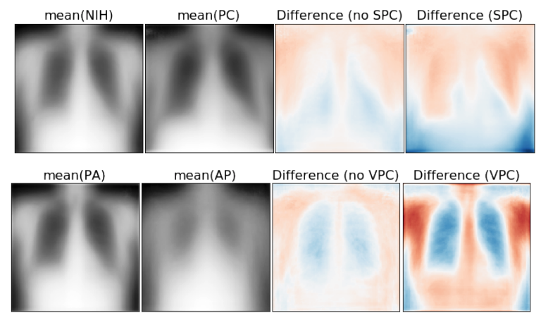
Poor generalization is one symptom of models that learn to predict target variables using spuriously-correlated image features present only in the training distribution instead of the true image features that denote a class. It is often thought that this can be diagnosed visually using saliency maps. We study if this is correct. In some prediction tasks, such as for medical images, one may have some images with masks drawn by a human expert, indicating a region of the image containing relevant information to make the prediction. We study multiple methods that take advantage of such auxiliary labels, by training networks to ignore distracting features which may be found outside of the region of interest, on the training images for which such masks are available. This mask information is only used during training and has an impact on generalization accuracy depending on the severity of the shift between the training and test distributions. Surprisingly, while these methods improve generalization performance in the presence of a covariate shift, there is no strong correspondence between the correction of saliency towards the features a human expert have labelled as important and generalization performance. These results suggest that the root cause of poor generalization may not always be spatially defined, and raise questions about the utility of masks as “attribution priors” as well as saliency for explainable predictions.
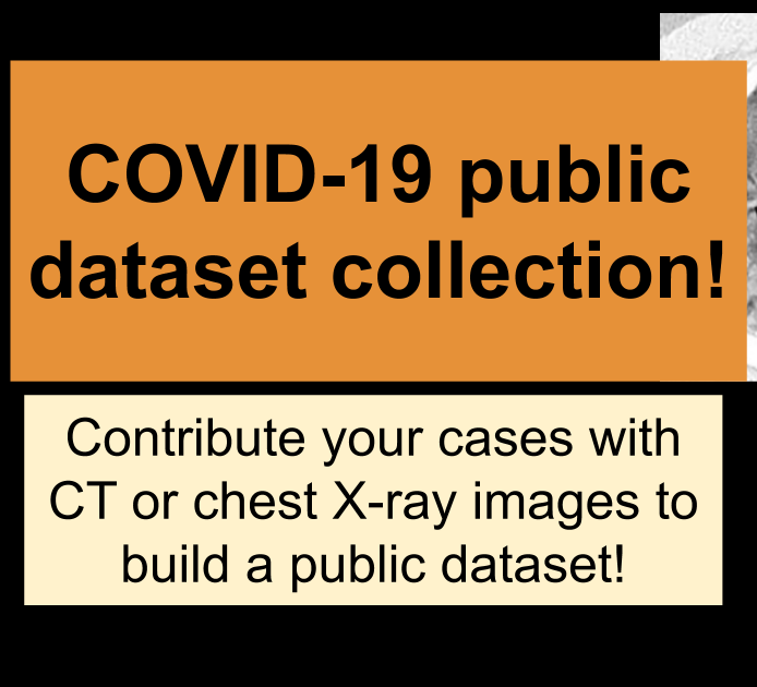
COVID-19 Image data collection
Project Summary: To build a public open dataset of chest X-ray and CT images of patients which are positive or suspected of COVID-19 or other viral and bacterial pneumonias (MERS, SARS, and ARDS.). Data will be collected from public sources as well as through indirect collection from hospitals and physicians. All images and data will be released publicly in this GitHub repo.

Hadrien Bertrand PhD
Mila

Lan Dao
Mila, Université de Montréal
Joseph Viviano
Mila
Vincent Frappier PhD
Paul Bertin
Mila, Université de Montréal
Joseph Paul Cohen PhD
Stanford University